Optical microscopy is a technique that allows the viewing of samples more closely using optical microscopes. It relies on light and one or more lenses to magnify samples. Optical microscopy is remarkably versatile, increasing the detail and contrast of a microscopic specimen. A range of applications rely on simple and complex microscopy techniques.
Fluorescence
Fluorescence microscopy is a type of optical microscopy that uses a fluorescent dye called fluorophores. When light hits the dye, it induces fluorescence rather than scattering or absorbing light, making tissue, cells, and proteins visible under a microscope. Fluorophores absorb energy from a specific wavelength known as the excitement range resulting in the energy’s re-emission in a wavelength known as the emission range.
Learn more about how we employ optical filters in fluorescence microscopy.
Phase Contrast

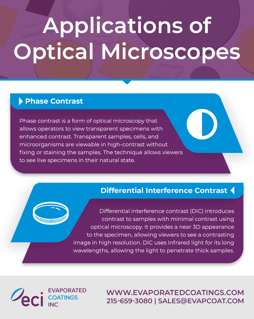
Click to expandPhase contrast is a form of optical microscopy that allows operators to view transparent specimens with enhanced contrast. Transparent samples, cells, and microorganisms are viewable in high-contrast without fixing or staining the samples. The technique allows viewers to see live specimens in their natural state.
Differential Interference Contrast
Differential interference contrast (DIC) introduces contrast to samples with minimal contrast using optical microscopy. It provides a near 3D appearance to the specimen, allowing viewers to see a contrasting image in high resolution. DIC uses infrared light for its long wavelengths, allowing the light to penetrate thick samples.
DIC creates a contrasting image when light passes through a polarizing filter and another polarized optical device. The polarized light passes through an objective-specific prism where the light beam is split and passes through a condenser. The condenser focuses the beams of light on specific points of the specimen.
The light beams pass through the specimen at various locations and various wavelengths. They move on to an objective lens that refocuses the beams on the rear of the focal plane. The nosepiece prism combines the beams, and the beam passes through the analyzer. The analyzer causes destructive and constructive interference, bringing the beams to the identical axis and plane. The light travels to the camera for the viewing of the DIC image.
Brightfield and Darkfield Illumination
Brightfield illumination presents a dark specimen on a bright background to create contrast. It is a simple technique that positions a light source below the sample. Light passes through the specimen to an objective lens and optical sensor. The darkness of the specimen increases with the specimen’s density. The more dense the sample is, the more pronounced the image will be.
Brightfield illumination is the result of these four key elements:
- Light Source
- Condenser Lens
- Objective Lens
- Eyepiece or Camera
Darkfield illumination creates a light specimen image on a dark background, contrasting the brightfield illumination technique. This technique enhances a specimen’s contrast without staining, allowing observation of living specimens.
Darkfield illumination begins with a light source that is obstructed by a dark field patch stop as it enters the microscope. The light is reduced to a ring where the condenser lens focuses it onto the sample. When the light hits the specimen, it transmits or scatters. The objective lens permits scattered light but blocks transmitted light with help from the dark illumination block.
Optical Microscopy Solutions From Evaporated Coatings
Optical microscopy improves specimen viewing by magnifying microscopic samples and enhancing their visibility with techniques and lenses. Fluorescence, phase contrast, brightfield illumination, darkfield illumination, and DIC allow optical microscopes to deliver a closer image in various applications.
At Evaporated Coatings, we specialize in high-quality optical coatings. We manufacture a range of optical filters, including excitement and emission filters for fluorescence microscopy. Our designers can help you find a cost-effective and high-performance solution, and our technicians use leading technology to manufacture the substrate and coatings you require. Contact us today to learn more about our high-quality optical coating solutions.
